Norepinephrine is a key neuromodulator that shapes brain states such as attention, stress, and learning. However, tracking its activity in the living brain remains challenging. A new study by Valentin Lu Rohner, Paul J. Lamothe-Molina, Zacharoula Kagiampaki, Tommaso Patriarchi and colleagues at UZH and collaborating institutions introduces next-generation fluorescent sensors that enable more sensitive and versatile imaging of norepinephrine signals. These tools allow researchers to visualize when and where this important neuromodulator is released, opening new possibilities for studying brain function in real time.
Norepinephrine plays a central role in regulating arousal, memory, and stress responses, yet capturing its dynamics across brain circuits has been technically difficult. Existing methods often lack either the spatial precision or the temporal resolution needed to follow rapid changes in neuromodulator release. To overcome these limitations, the researchers developed improved genetically encoded fluorescent indicators, nLightG2 and nLightR2, that emit signals when norepinephrine binds. They systematically tested these sensors across multiple experimental settings, from cultured cells and brain slices to live, behaving mice, demonstrating their ability to track norepinephrine dynamics across different levels of biological complexity.
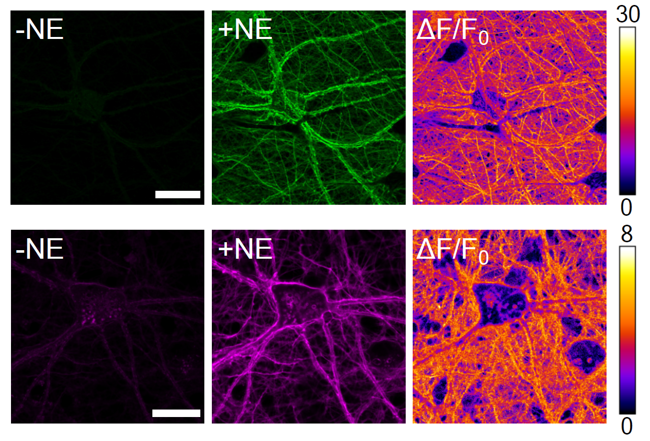
Testing these indicators across cells, brain tissue, and behaving animals showed a clear improvement over previous tools. The new sensors produced stronger and more reliable signals, enabling detection of even small, localized release events. For example, in the visual cortex of awake mice, the sensors revealed that norepinephrine is not released uniformly but in brief, spatially confined “microdomains” – small hotspots of activity that would previously have gone unnoticed. Similarly, during fear learning, the indicators captured how norepinephrine signals increase in specific brain regions in response to meaningful cues. These findings demonstrate that the new tools can track neuromodulator dynamics with high precision across different conditions.
By making it possible to watch norepinephrine signals unfold in real time, these advances provide a powerful new toolkit for studying how neuromodulators shape brain function. The improved sensitivity and flexibility of the sensors allow researchers to observe when and where these signals arise, and how they interact with neural activity across different brain regions and behavioral states. More broadly, this approach opens new ways to study how brain states emerge and change. It could also help uncover how disrupted neuromodulation contributes to brain disorders and inform more targeted treatments.
Reference: Rohner VL, Curreli S, Lamothe-Molina PJ, Kagiampaki Z, Yee AG, Nardin C, Eschholz L, Dieter A, Foustoukos G, Milanese P, Banterle L, Childs T, Canziani A, Dernic J, Ravotto L, Bhat MA, Sönmez L, Moreno Wasielewski L, Ziebarth T, Molnar MI, Huang G, Masseck OA, Weber B, Reiner A, Lüthi A, Ford CP, Wiegert JS, Ruediger S, Fellin T, Patriarchi T. Next-generation multicolor indicators for in vivo imaging of norepinephrine. Nature Methods. 2026. https://doi.org/10.1038/s41592-026-03006-z
Useful links:
Main image: The Patriachi Lab, University of Zurich
Representative images of the red (nLightR2, bottom) and green (nLightG2, above) norepinephrine sensors expressed in primary cortical rat neurons before and after the addition of norepinephrine (NE) and the pixel-wise heat map of the dynamic range.